
Summary
An emerging and highly promising class of biomarkers is based on extracellular vesicles (EVs). Liver-derived EVs carry cell-specific cargo, including proteins and microRNAs, that reflect hepatocyte-specific mechanisms such as metabolic stress, inflammation, and fibrogenesis. This cell-of-origin specificity enables superior measurement of steatosis and active MASH and supports longitudinal monitoring of disease activity. Furthermore, EV-based biomarkers have the potential to enable precision medicine approaches in drug development, including patient stratification and target-engagement assessment, thereby addressing a central unmet need in metabolic liver disease and MASLD/MASH therapeutic development.
At SBH Sciences, we are advancing a hepatocyte-isolated EVs platform designed to enable in-depth analysis of RNA and protein based biomarkers associated with NAFLD/NASH severity and progression, including pathways related to inflammation, lipid stress, apoptosis, and fibrosis. This approach supports a comprehensive and dynamic assessment of disease biology and treatment response. We are excited to collaborate with academic and industry partners to further develop and apply this platform in preclinical and translational research to accelerate the development of disease-modifying therapies for liver disease.
Potential unmet diagnostic need in MASLD/MASH clinical studies:
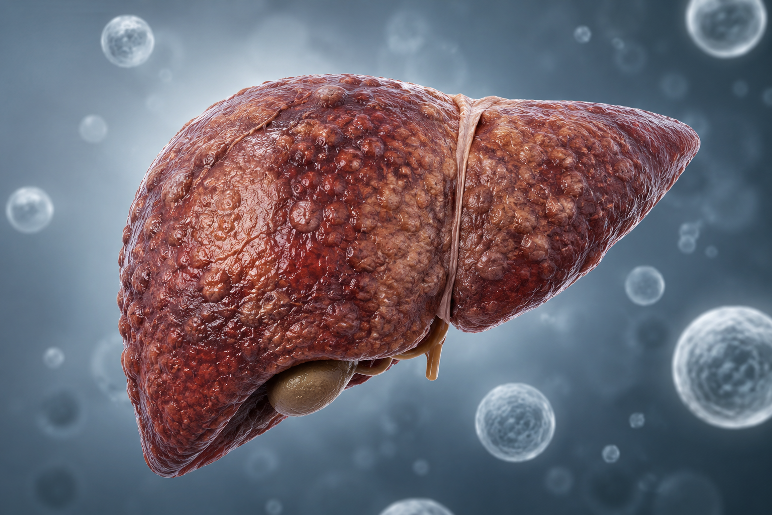
Metabolic dysfunction associated steatotic liver disease (MASLD) is the most prevalent chronic liver disease worldwide and a leading cause of liver-related morbidity and mortality. Its progressive inflammatory subtype, metabolic dysfunction–associated steatohepatitis (MASH), drives fibrosis, cirrhosis, hepatocellular carcinoma, and liver transplantation. While significant progress has been made in advancing therapeutic strategies, existing diagnostic technologies to accurately assess mechanistic drug effects and monitor treatment response remain a major bottleneck in MASH clinical development (Younossi et al., 2018; Rinella et al., 2023; Sanyal et al., 2021).
Extracellular Vesicles as a Novel Diagnostic Platform
Extracellular vesicles (EVs) are membrane-enclosed nanoparticles released by almost all cell types under physiological and pathological conditions. EV cargo includes proteins, lipids, and nucleic acids that reflect the cellular status of their parent cell and provide a strategy to interrogate cellular pathways from blood samples. EVs are stable in circulation, making them attractive liquid biopsy substrates (Raposo & Stoorvogel, 2013; Théry et al., 2018).
In liver disease, EVs have emerged both as mediators of pathogenesis and as circulating reporters of hepatocellular stress, inflammation, and fibrosis. In MASLD/MASH, hepatocytes, hepatic stellate cells, Kupffer cells, liver sinusoidal endothelial cells, and infiltrating immune cells increase EV release in response to lipotoxicity, oxidative stress, and inflammatory signaling. These EVs enter the circulation and can be sampled non-invasively, providing a unique window into intrahepatic biology not captured by conventional plasma biomarkers (Eguchi et al., 2017; Povero et al., 2014; Hirsova et al., 2016).
Utility of EVs Based Biomarkers in MASLD/MASH
Multiple studies demonstrate that circulating EV profiles are altered in MASLD and MASH, with EV abundance correlating with disease severity, inflammatory activity, and fibrosis stage. However, EV quantity alone lacks specificity due to contributions from non-hepatic tissues in systemic metabolic disease. Recent advances therefore focus on cell-of-origin–resolved EV profiling and molecular cargo analysis to improve specificity and diagnostic performance (Welton et al., 2017; Kornek et al., 2012).
Liver-Derived EVs
Hepatocyte-derived EVs can be isolated using surface markers that identify liver origin. Markers such as asialoglycoprotein receptor-1 (ASGR1) significantly enhance diagnostic resolution by enriching liver-specific signals and reducing background from platelets and immune cells. Liver-derived EVs show strong associations with steatohepatitis activity and fibrosis severity and outperform total plasma EV measurements. Importantly, EV cargo analytes from hepatocyte-specific fractions more accurately reflect intrahepatic pathology than generic circulating biomarkers (Kowal et al., 2016; Hirsova & Ibrahim, 2021).
EVs Cargo MicroRNAs and Protein Biomarkers
EV-associated microRNAs (miRNAs) represent the most mature class of EV-based biomarkers in MASLD/MASH. Liver-enriched miRNAs such as miR-122, along with inflammation- and apoptosis-associated miRNAs including miR-34a, miR-192, and miR-21, are consistently elevated in EVs from patients with steatohepatitis and correlate with disease severity. Multivariate EV miRNA panels have demonstrated improved discrimination between simple steatosis and MASH compared with individual markers (Pirola et al., 2015; Cheung et al., 2008; Cermelli et al., 2011).
In parallel, proteomic profiling of EVs has identified candidate protein biomarkers involved in inflammation, adhesion, and fibrogenic signaling, including integrins and hepatocyte stress–associated proteins. Although EV proteomics is less mature than miRNA analysis, these protein signatures offer strong translational potential as assay technologies become increasingly standardized and scalable (Povero et al., 2014; Hirsova et al., 2016).
These attributes position EV-based assays as a transformative tool for precision diagnostics and clinical trial optimization in MASLD/MASH.
SBH Sciences diagnostic panel to assess NAFLD/NASH severity and disease progression
We have identified a panel of candidate RNA and protein biomarkers associated with NAFLD/NASH severity and disease progression, including markers of inflammation, lipid stress, apoptosis, and fibrosis. The proposed panel includes the analytes listed in Table 1 (protein biomarkers) and Table 2 (miRNA biomarkers).


SBH Sciences is advancing a hepatocyte-isolated EV platform designed to enable the discovery and analysis of novel RNA and protein biomarkers associated with NAFLD/MASH severity and progression, including key pathways such as inflammation, lipid stress, apoptosis, and fibrosis. Our platform is built to support precision medicine by enabling sensitive, mechanism driven biomarker readouts that can inform patient stratification, target engagement, and treatment response. This comprehensive and dynamic assessment of liver pathology positions our technology as a powerful tool for both biomarker discovery and clinical development.
We welcome collaborations with academic and industry partners to further develop and apply this platform in preclinical and translational research to support the development of disease-modifying therapies for liver disease.
Ready to accelerate your liver research? Connect with SBH Sciences. mstewart@sbhsciences.com | www.sbhsciences.com/metabolic
References
Younossi ZM et al. (2018). Global epidemiology of NAFLD. Hepatology.
Rinella ME et al. (2023). AASLD nomenclature update: MASLD/MASH. Hepatology.
Sanyal AJ et al. (2021). Challenges and opportunities in NASH drug development. Gastroenterology.
Raposo G, Stoorvogel W. (2013). Extracellular vesicles: exosomes, microvesicles, and friends. J Cell Biol.
Théry C et al. (2018). Minimal information for studies of extracellular vesicles (MISEV2018). J Extracell Vesicles.
Eguchi A et al. (2017). Extracellular vesicles in liver disease. Hepatology.
Povero D et al. (2014). Lipotoxic hepatocyte-derived EVs. Hepatology.
Hirsova P et al. (2016). Lipotoxic EV signaling in NASH. Hepatology.
Welton JL et al. (2017). EV profiling in disease. J Extracell Vesicles.
Kornek M et al. (2012). Microparticles in liver disease. Hepatology.
Kowal J et al. (2016). EV heterogeneity and markers. PNAS.
Hirsova P, Ibrahim SH. (2021). EVs as biomarkers in liver disease. Nat Rev Gastroenterol Hepatol.
Pirola CJ et al. (2015). Circulating miRNAs in NAFLD. Gut.
Cheung O et al. (2008). miR-122 in liver disease. Hepatology.
Cermelli S et al. (2011). Circulating miRNAs as biomarkers. PNAS.
Ready to add NF-L1 to your biomarker strategy? Contact SBH Sciences to learn more.
mstewart@sbhscience.com | www.sbhsciences.com