
SBH Western Blot Capabilities
At SBH Sciences, we offer a next generation Simple Western™ Blot CRO service using our in house ProteinSimple instruments, the WES, Jess and the PeggySue, which are designed to replace and dramatically improve traditional western blotting workflows.
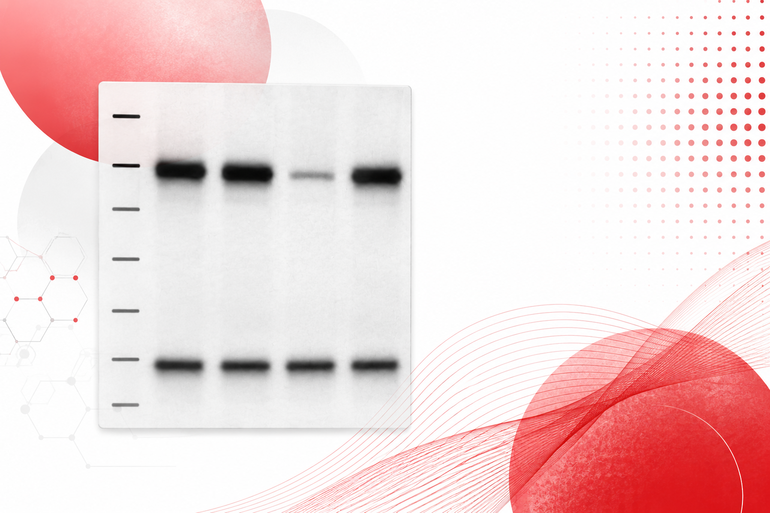
Simple Western™ is an innovative capillary electrophoresis (CE) based immunodetection platform that combines the high sensitivity and antibody specificity of traditional western blotting with the high resolution and reproducibility of capillary electrophoresis — all in a single, fully automated workflow.

Proteins can be separated either by size (as in traditional SDS-PAGE/Western) or by isoelectric point (pI), as in isoelectric focusing (IEF). This dual-mode capability gives researchers flexibility across a wide range of protein characterization applications.
Our analytical capabilities include everything from protein quantification and characterization to immunoassay multiplex protein detection and kinase inhibitor target profiling.

Key Advantages Over Traditional Western Blotting
- Quantitative, reproducible protein analysis with coefficients of variation (CVs) under 10%
- High-throughput capability — up to 96 samples analyzed per run
- Ability to multiplex and normalize to standard loading control proteins
- Both size-based (MW) and charge-based (pI/IEF) separation modes
- Fully automated workflow reduces operator variability and hands-on time
- Compatible with precious or limited clinical samples (e.g., fine needle aspirates, serum)
- Electropherogram view provides quantitative peak data alongside traditional blot visualization

Real World Use Cases
Below are a selection of case studies that demonstrate the breadth and depth of analyses supported by SBH Sciences’ Simple Western™ service. All experiments were performed on the Wes and/or PeggySue platforms in our laboratory.
Case Study 1: Drug Effect on Protein Phosphorylation - Charge (IEF) Analysis
Problem: How do you quickly determine if drug is activating or suppressing a protein signaling pathway and how do you track that response through the changes in living cells?
Platform: PeggySue (IEF/Charge Based)
Sample Type: cell culture lysate
Time Points: 0, 15, 30, 60, 120, 240 minutes
Two chemotherapeutic drugs were tested at a single concentration against cell culture cells across a time course of 0 to 240 minutes. Charge-based (IEF) Simple Western analysis revealed dramatically different effects on protein phosphorylation:
- Drug A caused a progressive decrease in protein phosphorylation over time, observable as a shift in pI toward higher values (less negatively charged isoforms) from the 15-minute time point onward.
- Drug B caused a progressive increase in protein phosphorylation over time, with bands shifting toward lower pI values (more negatively charged isoforms) as treatment continued.

Results were visualized in Standard Western View across the pI range of 4–8, with the phosphorylation state clearly resolved at each time point.
Case Study 2: Characterization of Biotherapeutic Production - Post-Translational Modification Analysis by Charge (IEF)
Problem: How do you detect and quantify subtle differences in post-translational modifications between biotherapeutic production lots before they become a quality or regulatory problem?
Platform: PeggySue (IEF/Charge Based)
Sample Type: Biotherapeutic production Lot #1 vs Lot #2
Two production lots of a biotherapeutic protein were analyzed side-by-side using IEF-based Simple Western. The electropherogram view revealed distinct charge variant profiles between Lot #1 and Lot #2 across a pI range of approximately 4–9.
Individual peaks were resolved and quantitatively measured at the following pI values: 5.06, 5.16, 5.26, 5.37, 5.47, 5.57, 5.69, 5.82, and 6.11. The relative abundance of each charge variant differed significantly between the two lots, with Lot #1 showing higher signal at later (more basic) pI positions and Lot #2 showing more signal in early (more acidic) positions.
This level of quantitative resolution between individual charge variants is not achievable by traditional gel-based western blotting, demonstrating the superior capability of Simple Western for biotherapeutic lot release and comparability testing.
Case Study 3: Drug Response Measurement in Animal Tumors - Fine Needle Aspirate Analysis — Time Course
Problem: How do you measure a drug’s molecular effect inside a living tumor using only the tiny precious sample volume available from a fine needle aspirate?
Platform: PeggySue (IEF/Charge Based)
Sample Type: Fine needle aspirates from animal tumors
Time Points: T0(pre-dose), T2, T8, T24 hours post injection
A drug was administered at time zero and tumor cells were extracted via Fine Needle Aspirate at 2, 8, and 24 hours post-injection. Both Standard Western View and Electropherogram View were generated for each time point.
Results demonstrated significant and progressive increases in protein phosphorylation from T2 through T24 hours, clearly visible as emergence and growth of lower-pI peaks in the electropherogram. The T0 (pre-dose) sample showed minimal phosphorylated species, establishing a clean baseline for comparison.
This application highlights the platform’s suitability for precious, low-volume clinical and preclinical samples — a setting where traditional western blotting is often not feasible due to sample quantity constraints.
Case Study 4: Identification of Clinical Trial Patient Response - Protein Quantification from Human Serum Samples
Problem: How do you rapidly screen dozens of clinical trial patients to identify who is actually responding to treatment, accurately and consistently across all samples in a single run?
Platform: PeggySue (size-based)
Sample Type: Human serum
Sample Count: 24 patient sera, each diluted 1:10, 1:20, and 1:40
Throughput: Up to 96 samples analyzed per run
Twenty-four clinical trial patient sera were analyzed at three dilutions (1:10, 1:20, 1:40) simultaneously in a single run. Antibody reactivity was measured and the area under the major peak was quantified for each sample.

Of the 24 patients analyzed, 8 showed a significant decrease in protein level, indicating a drug response. The remaining 16 patients showed protein levels consistent with non-responders. This stratification was achieved rapidly and quantitatively across all samples in a single automated run.
This use case demonstrates the platform’s value in translational and clinical research, enabling high-throughput biomarker-based patient stratification with the consistency required for clinical data.
Case Study 5: Monoclonal Antibody Selection - Biomarker Identification Across Multiple Cell Lines
Problem: How do you efficiently narrow down multiple antibody candidates across multiple cell lines to find the one that reliably detects your target protein without running weeks of individual gel experiments?
Platform: Wes (size-based)
Antibodies Tested: 5 monoclonal antibodies (Antibody 1–5)
Cell Lines: MM1S, AML5, MNF560, NB2-11, R&D TF1, MPC-11
Six different cell lines were analyzed simultaneously against each of five monoclonal antibody candidates. Beta-Actin was used as a loading control to normalize expression levels across samples. All 30 sample-antibody combinations (6 cell lines × 5 antibodies) were evaluated in a single experimental run.
Key Findings
- Antibody 4 demonstrated the best and most consistent reactivity to the target biomarker across all six cell lines, making it the leading candidate for downstream assay development.
- Antibody 5 showed reactivity to a protein fragment rather than the full-length target protein, flagging it as unsuitable for the intended application.
- Antibodies 1, 2, and 3 showed variable or weak reactivity across cell lines.
Running 6 cell lines × 5 antibodies in a single automated run would require multiple gel runs and days of manual work with traditional western blotting — here it was accomplished in a single automated session.

Case Study 6: Drug Response Time Course — Washout Analysis - Determining Duration of Drug Effect
Problem: How do you determine how long a drug’s effect on a target biomarker protein persists after the drug is removed?
Platform: Wes (size-based)
Drug Concentrations: 0, 6.25, 12.5, 25, 50 nM
Time Points: Day 1 (with drug), Day 2 (washout), Day 3 (washout)
Loading Control: GAPDH
Drug was applied at four concentrations on Day 1 and then removed (washed out) after 24 hours. Cells were analyzed for biomarker protein expression on Day 1 (with drug), Day 2 (one day post-washout), and Day 3 (two days post-washout). GAPDH was used as a loading control for normalization.
Results
- Day 1: Significant concentration-dependent decrease in biomarker protein expression observed at all active drug concentrations compared to the 0 nM control.
- Day 2 (post-washout): Biomarker protein remained significantly suppressed despite drug removal, indicating the drug’s effect persisted through Day 2.
- Day 3 (post-washout): Biomarker protein expression recovered toward baseline levels across all concentration groups.

Conclusion: The drug effect on the target biomarker lasts approximately two days, providing a clear pharmacodynamic window for dosing schedule design.

Case Study 7: Calculating Concentration of an Unknown Biomarker - Quantification Using a Known Protein Standard Curve
Problem: How do you get beyond a yes/no blot result and get an actual protein concentration from an unknown sample with enough statistical rigor to support downstream decisions?
Platform: Wes (size-based)
Standard Dilutions: 100%, 80%, 60%, 40%, 20% of known protein
Unknowns: 3 unknown samples (all run in triplicate)
A five-point dilution series of a known protein was run in the same capillary array as three unknown samples, all in triplicate. Average area values were calculated for each known dilution point and plotted against concentration to generate a standard curve.
Unknown protein concentrations were interpolated from the slope of the dilution curve using chemiluminescent area values. The approach demonstrated that Simple Western can be used as a quantitative immunoassay — not just a qualitative blot — providing absolute protein concentration estimates from unknown samples.
 Running all samples (standard curve + 3 unknowns in triplicate = 14 total samples) in a single automated run ensures consistency across the standard and unknowns, removing the inter-run variability that affects multi-gel western workflows.

Case Study 8: Optimization of Biomarker Recovery from Clinical Samples - Elution Efficiency Analysis by Size
Problem: How do you know whether your sample preparation protocol is actually recovering your target biomarker and how much you lose with each elution step?
Platform: Wes (size-based)
Design: 10 sequential eluate fractions + 10 matched input samples
Input CV: 3.8% (Avg. Area: 437, SD: 16)
A five-point dilution series of a known protein was run in the same capillary array as three unknown samples, all in triplicate. Average area values were calculated for each known dilution point and plotted against concentration to generate a standard curve.
The Eluate/Input Ratio was tracked across all 10 elution fractions. Results showed:
- Elution 1 yielded the highest recovery (~11% of input), with each subsequent elution recovering progressively less material.
- By Eluate 9, recovery approached 0%, indicating near-complete recovery across the combined fractions.
- The electropherogram confirmed the MW of the target biomarker at approximately 116 kDa across eluate fractions 1, 3, 5, 7, and 9.

This data supports the use of multiple sequential elutions for maximum biomarker recovery in clinical sample processing workflows.

Case Study 9: Knockdown Experiment Using siRNA - Validation of Target Gene Silencing
Problem: How do you confirm that your siRNA is specifically and durably silencing its target protein (and not producing off-target effects) at multiple time points post-transfection?
Platform: Wes (size-based)
Conditions: (1) Untransfected, (2) Non-Targeted Control siRNA, (3) Targeted siRNA
Time Points: 24 hours and 72 hours post-transfection
Three conditions (untransfected, NTC siRNA, targeted siRNA) were analyzed at 24- and 72-hour time points. Both Standard Western View and Electropherogram View were generated. A loading control was run in parallel to normalize expression levels.
Results clearly demonstrated effective knockdown of the targeted protein:
- At 24 hours: The targeted siRNA condition showed dramatically reduced peak signal compared to untransfected and NTC controls, which were comparable to each other.
- At 72 hours: Knockdown was maintained, with the targeted siRNA condition continuing to show significantly reduced protein expression.
- NTC siRNA results were consistent with untransfected controls at both time points, confirming the knockdown was sequence-specific.

Interested in how automated western blot technologies could help advance your drug discovery pipeline? To discuss your project requirements and receive a custom proposal, please contact us: anir@sbhsciences.com
At SBH Sciences, we support biotech and pharmaceutical partners with cell-based assays, biomarker testing, assay validation, and translational research services designed to accelerate discovery through clinical development.