
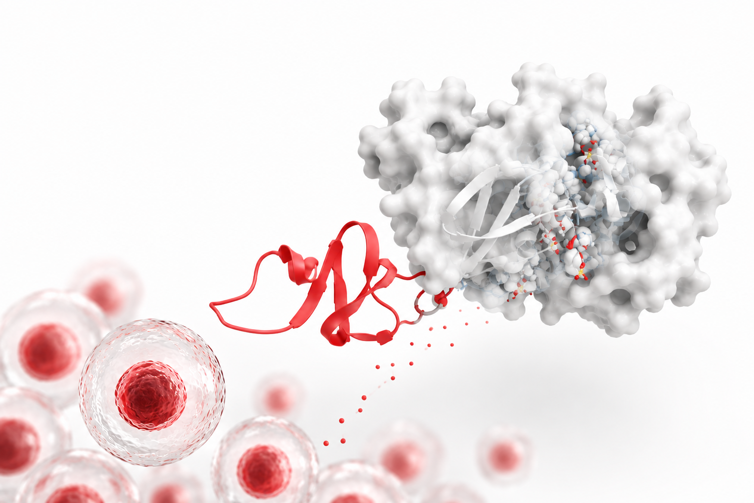
Activin A has become one of the most widely used growth factors in modern stem cell biology. Originally identified for its role in reproductive endocrinology, this TGF-β superfamily protein is now a cornerstone reagent for guiding pluripotent stem cells toward specific fates, and a growth factor that SBH Sciences manufactures as a high-purity recombinant protein for exactly these kinds of applications.
What is Activin A?
Activin A is a 26 kDa disulfide-linked homodimer made up of two beta-A chains, each containing 116 amino acid residues, and it belongs to the TGF-β superfamily of growth and differentiation factors. It signals primarily through the SMAD2/3 pathway after binding to type I and type II activin receptor complexes, triggering a wide range of downstream effects on cell proliferation, differentiation, and survival [2]. Its activity can be neutralized by inhibins and by the diffusible antagonist follistatin, giving researchers a natural "off switch" when designing experiments.
Because of this broad signaling reach, Activin A shows up in research spanning stem cell biology, oncology, immunology, reproductive medicine, metabolism, and regenerative medicine.
Activin A in Stem Cell Research and Differentiation
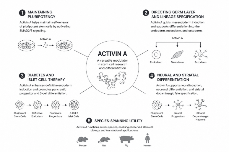
It's in pluripotent stem cell biology that Activin A has become nearly indispensable. Its effects are highly dependent on concentration, timing, and co-factors, allowing the same protein to maintain stem cells in an undifferentiated state in one context and drive them toward a mature, specialized cell type in another.
Maintaining pluripotency: Activin A is one of the key soluble factors that allows human embryonic stem cells to be maintained in an undifferentiated state without feeder layers, supporting long-term self-renewal while preserving normal karyotype and pluripotency markers.
Directing germ layer and lineage specification: Activin A is a primary driver of definitive endoderm formation, the first step in generating pancreatic, hepatic, and intestinal cell types from human pluripotent stem cells [3]. One research group found that human embryonic stem cells shift toward endoderm as Activin A concentration rises relative to BMP4 and VEGF, with each of the three cytokines steering differentiation toward a different lineage [4]. Depending on concentration, timing, and the presence of co-factors like BMP4 and WNT modulators, Activin A also contributes to cardiac mesoderm induction during cardiomyocyte, endothelial, and smooth muscle cell differentiation protocols [3]. Further downstream, Activin A can be added to definitive endoderm cells to induce hepatic or pancreatic progenitor fates by mediating the Nodal signaling pathway; combining Activin A with LiCl-driven Wnt activation increases the efficiency of generating these endoderm-derived progenitors [5]. A separate line of research has shown that Activin A, together with inhibition of BMP and Wnt signaling, can direct mouse embryonic stem cells toward Noto-expressing, notochord-like cells [6].
Skeletal lineage differentiation: Activin A also guides mesenchymal and skeletal fates. In combination with BMP4, TGF-β3, and Wnt3a, it has been used to push embryonic stem cells through chondrogenic induction, with long-term Activin A supplementation promoting the formation of articular cartilage [7]. In mesenchymal progenitor cells, Activin A is required for chondrogenic and osteogenic differentiation: both absolute Activin A levels and the Activin A-to-follistatin ratio shape that differentiation, and Activin A separately inhibits progenitor cells from committing to fat (adipogenesis) [8].
Diabetes and islet cell therapy: One of the most clinically significant applications today is in generating insulin-producing islet cells from induced pluripotent stem cells (iPSCs) for diabetes therapy. GMP-grade Activin A is used at the very first differentiation step, driving iPSCs toward definitive endoderm, in manufacturing processes now being scaled for cell therapies such as Vertex's VX-880, which has advanced to Phase III trials [9]. Efficient, reproducible, high-quality Activin A is a rate-limiting ingredient in making these large-scale differentiation protocols commercially viable. A related, earlier line of work found that gnotobiotic porcine skin-derived stem cells treated with Activin A, glucagon-like peptide 1 (GLP-1), and nicotinamide could be converted into insulin-producing cells that expressed insulin and increased insulin synthesis in response to glucose stimulation, pointing to alternative non-pancreatic starting material for beta-cell-like therapies [10].
Neural and striatal differentiation: Activin A also plays a role in neuronal subtype-specific differentiation, including directing human pluripotent stem cells toward striatal medium spiny neuron fates relevant to modeling and potentially treating Huntington's disease.
Species-spanning utility: This isn't limited to human cells, either. Activin A also regulates pluripotency-associated gene expression (such as NANOG and SOX2) in bovine embryonic stem cells, underscoring how conserved this signaling axis is across mammalian species and how useful it is for veterinary and agricultural stem cell research alongside human applications [11].
A Multifunctional Cytokine Beyond Stem Cells
Given how many cell types and pathways Activin A touches, it's no surprise that its influence extends well past the stem cell field.
Cancer: a context-dependent role: Elevated Activin A has been linked to poor prognosis across several tumor types, where it can promote cell growth, migration, angiogenesis, and immune evasion, making it an emerging therapeutic target [2]. Yet much of the earliest work on Activin A in cancer actually found the opposite effect. Activin A halts human breast cancer cells in the G1 phase of the cell cycle through the p38 MAPK pathway [12,13], and it inhibits proliferation of human colon cancer (RKO) cells by inducing SLC5A8 expression through SMAD3 signaling [14]. It has also been shown to inhibit growth in prostate, leukemia, vascular endothelial, fetal adrenal, and vascular smooth muscle cells, though not always through the same mechanism [15]. Other tumor contexts flip the script: elevated Activin A in glioblastoma produces a dose-dependent increase in DNA synthesis and enhances proliferation of U87 glioblastoma cells [16], and in multiple myeloma, tumor cells drive marrow stromal cells to secrete more Activin A, which suppresses osteoblast differentiation and helps create a bone microenvironment favorable to the cancer; blocking Activin A reverses that osteoblast suppression and slows tumor growth in humanized mouse models [17]. A related study in osteoblast-like cells found that Activin A shifts cells toward the G1 phase and reduces cell death overall, without increasing proliferation, suggesting the added cell numbers come from reduced turnover rather than faster division [18]. This dual, tumor-type-specific behavior is part of why Activin A is being pursued as a drug target: as of mid-2011, Acceleron Pharma and Celgene were running a Phase 2/3 trial of ACE-011 (sotatercept), a soluble receptor fusion protein with high affinity for Activin A, for chemotherapy-induced anemia in metastatic non-small cell lung cancer.
Immune regulation and inflammation: Recent work has shown that Activin A activates a SMAD3-dependent pathway in macrophages that acts as a natural brake on inflammation during sepsis and psoriasis in mouse models [19]. That anti-inflammatory brake sits alongside older findings that neutrophils are themselves a significant source of Activin A, with TNF-α driving neutrophils to secrete Activin A within an hour of stimulation, well before Activin mRNA expression rises [20]. Serum Activin A is also elevated in several rheumatic diseases: levels run higher in rheumatoid arthritis, systemic lupus erythematosus, and osteoarthritis than in healthy controls, with rheumatoid arthritis and lupus levels significantly higher than osteoarthritis and a positive correlation between Activin A and disease activity in both [21]. In the airway, blocking Activin A with an antibody prevents the IL-25 increase seen in asthma models, limiting airway hyper-reactivity, remodeling, and collagen deposition, making it a candidate asthma target as well [22].
Reproductive biology and pregnancy: In pregnancy biology, Activin A regulates trophoblast invasion through a SOX4 and microRNA-103a-3p network, with elevated levels associated with preeclampsia [23]. That mechanistic picture lines up with earlier clinical observations: serum Activin A, alongside Inhibin A and decreased placental growth factor (PlGF), can help predict preeclampsia [24]. Activin A is also essential to male fertility; levels rise in the testis (but not the ovary) shortly after sex determination, and mice lacking Activin A have smaller testes and fewer Sertoli cells, since Activin A dosage sets the balance between Sertoli and germ cell numbers [25]. Separately, Activin A produced by fetal Leydig cells is required for fetal testis development [26]. On the maternal side, Activin A reduces production of cell adhesion molecules in a way that may explain implantation failure in endometriosis-associated infertility [27], while serum Activin A tends to run lower in ectopic pregnancies than in normal intrauterine pregnancies, a difference that can help flag ectopic pregnancy [28], especially when combined with progesterone, VEGF, and Inhibin A into a four-marker panel [29]. Activin A additionally inhibits activation of human primordial follicles in a dose-dependent manner, tied to its dose-dependent promotion of granulosa cell proliferation [30].
Musculoskeletal and metabolic roles: In spaceflight physiology, blocking the myostatin/Activin A signaling pathway protected mice from the muscle and bone loss typically seen during extended time in microgravity, a finding with implications for astronaut health and, more broadly, for disuse atrophy on Earth [31]. On Earth, inhibiting Activin A signaling stimulates bone formation and prevents cancer-induced bone damage without affecting tumor growth or proliferation, separating its skeletal effects from its tumor effects [32]. Activin A also drives fibrosis: it activates the ALK4-SMAD pathway in systemic sclerosis, where serum Activin A and Activin receptor type 1B expression both run higher than in healthy controls, and Activin A directly induces collagen production, making the Activin A-ALK4-SMAD axis a candidate treatment target [33]. In the pancreas, Activin A shifts beta cells toward a more immature, faster-proliferating state with reduced insulin expression, an effect reversed by exogenous follistatin [34]. And in adipose tissue, Activin A regulates the pool of undifferentiated progenitor cells and is expressed at higher levels in obese patients than in lean subjects [35].
Other systems: liver, neurons, heart, and wound healing: In the liver, older rat models show a route toward better transplant outcomes: fetal liver stem/progenitor cells repopulate the livers of older rats 4- to 5-fold more effectively than younger rats, because older rats have fewer Activin receptors and thus more resistance to Activin A's inhibition of adult hepatocyte proliferation, a combination of limited hepatocyte proliferation and increased apoptosis that favors the transplanted cells [36]. In neurons, Activin A promotes differentiation and supports survival independent of the usual SMAD pathway, instead acting through the Activin type 1 receptor [37]. In the heart, a sheep model shows that Activin A released during cardiopulmonary bypass is biologically active and reaches higher levels than surgery alone would produce, tying it to the adverse outcomes of systemic inflammation after cardiac surgery [38]; separately, Activin A levels track with carcinoid heart disease severity and independently predict its presence [39]. More broadly, Activin A is understood to play a role in inflammation, tissue repair, and cytoprotection across organs, accelerating wound healing and drawing interest as a potential neuroprotective agent [40].
Why Assay Quality Matters
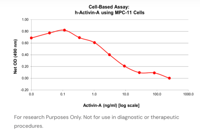
Given how concentration-dependent and context-dependent Activin A's effects are, the same protein maintains pluripotency at one dose and drives definitive endoderm differentiation at another, reliable, well-characterized recombinant protein is critical. SBH Sciences produces its recombinant human Activin A in Hi5 insect cells as a soluble 26 kDa homodimer corresponding to amino acid residues 311–426 of the full-length protein. Potency is validated using a cell-based bioassay, measuring the protein's ability to inhibit proliferation of the murine MPC-11 cell line, with an expected ED50 in the range of 0.5–2.0 ng/mL [1]. This kind of rigorous, batch-to-batch functional testing is what allows researchers to trust that a given lot will perform consistently in a differentiation protocol, whether that protocol is running in an academic lab or a GMP manufacturing suite.
Partnering With SBH Sciences
SBH Sciences is looking for partners to investigate its recombinant Activin A as both a therapeutic agent and a diagnostic tool. Our extensive bioassay support, paired with highly pure protein, is also well suited to projects aimed at developing anti-Activin A therapeutics, whether small-molecule drugs or monoclonal antibodies. We're always open to new collaborations. We’d love to hear from you.
References
- SBH Sciences. "Human Activin A Recombinant Protein." SBH Sciences Blog. https://sbhsciences.com/blog/human-activin-a-recombinant-protein
- Ries, A., Schelch, K., Falch, D., Pany, L., Hoda, M.A., & Grusch, M. (2020). Activin A: an emerging target for improving cancer treatment? Expert Opinion on Therapeutic Targets, 24(10), 985–996. https://doi.org/10.1080/14728222.2020.1799350
- Gao, Y., & Pu, J. (2021). Differentiation and Application of Human Pluripotent Stem Cells Derived Cardiovascular Cells for Treatment of Heart Diseases: Promises and Challenges. Frontiers in Cell and Developmental Biology, 9, 658088. https://www.frontiersin.org/journals/cell-and-developmental-biology/articles/10.3389/fcell.2021.658088/full
- Outten, J.T., et al. (2011). Stem Cell Research, 6(2), 129–142.
- Li, F., et al. (2011). Journal of Cellular Biochemistry, 112(4), 1022–1034.
- Winzi, M., et al. (2011). Stem Cells and Development.
- Waese, E.Y., et al. (2011). Stem Cell Research, 6(1), 34–49.
- Djouad, F., et al. (2010). Stem Cell Research & Therapy, 1(2), 11.
- ACROBiosystems. "Enabling large-scale iPSC-derived islet differentiation with GMP activin A and KGF." News-Medical. https://www.news-medical.net/whitepaper/20260122/Enabling-large-scale-iPSC-derived-islet-differentiation-with-GMP-activin-A-and-KGF.aspx
- Yang, J.H., et al. (2010). Biochemical and Biophysical Research Communications, 397(4), 679–684.
- Soto, D.A., Navarro, M., Zheng, C., Halstead, M.M., et al. (2021). Simplification of culture conditions and feeder-free expansion of bovine embryonic stem cells. Scientific Reports, 11. https://www.nature.com/articles/s41598-021-90422-0
- Burdette, J.E., et al. (2005). Cancer Research, 65(17), 7968–7975.
- Cocolakis, E., et al. (2001). The Journal of Biological Chemistry, 276(21), 18430–18436.
- Zhang, Y., et al. (2010). The International Journal of Biochemistry & Cell Biology, 42(12), 1964–1972.
- Chen, Y.G., et al. (2002). Experimental Biology and Medicine (Maywood, NJ), 227(2), 75–87.
- Zhang, D.F., et al. (2010). The Journal of International Medical Research, 38(4), 1343–1353.
- Vallet, S., et al. (2010). Proceedings of the National Academy of Sciences of the United States of America, 107(11), 5124–5129.
- Rosenberg, N., et al. (2010). Experimental and Clinical Endocrinology & Diabetes, 118(10), 708–712.
- Gauthier, T., Lim, Y.J., Jin, W., et al. (2025). Activin A activation of Smad3 mitigates innate inflammation in mouse models of psoriasis and sepsis. Journal of Clinical Investigation, 135(9), e187063. https://www.jci.org/articles/view/187063
- Chen, Y., et al. (2011). Immunology and Cell Biology.
- El-Gendi, S.S., et al. (2010). International Journal of Rheumatic Diseases, 13(3), 273–279.
- Gregory, L.G., et al. (2010). American Journal of Respiratory and Critical Care Medicine, 182(2), 143–154.
- Xie, J., Shannon, M.J., Zhu, H., Dong, M., Beristain, A.G., & Leung, P.C.K. (2026). Elucidating the roles of microRNA-103a-3p in trophoblast invasion and SOX4-mediated extravillous differentiation induced by activin A. Cell Death & Disease, 17, 466. https://www.nature.com/articles/s41419-026-08665-6
- Yu, J., et al. (2011). Ultrasound in Obstetrics & Gynecology, 37(5), 528–533.
- Mendis, S.H., et al. (2011). Biology of Reproduction, 84(2), 379–391.
- Archambeault, D.R., et al. (2010). Proceedings of the National Academy of Sciences of the United States of America, 107(23), 10526–10531.
- Stoikos, C.J., et al. (2010). Human Reproduction, 25(7), 1767–1774.
- Florio, P., et al. (2011). Gynecological Endocrinology, 27(6), 391–395.
- Rausch, M.E., et al. (2011). Obstetrics and Gynecology, 117(3), 573–582.
- Ding, C.C., et al. (2010). Journal of Assisted Reproduction and Genetics, 27(4), 141–147.
- Lee, S.J., Lehar, A., Meir, J.U., et al. (2020). Targeting myostatin/activin A protects against skeletal muscle and bone loss during spaceflight. Proceedings of the National Academy of Sciences, 117(38), 23942–23951. https://www.pnas.org/doi/10.1073/pnas.2014716117
- Chantry, A.D., et al. (2010). Journal of Bone and Mineral Research, 25(12), 2357–2370.
- Takagi, K., et al. (2011). Journal of Autoimmunity, 36(3–4), 181–188.
- Szabat, M., et al. (2010). Diabetologia, 53(8), 1680–1689.
- Zaragosi, L.E., et al. (2010). Diabetes, 59(10), 2513–2521.
- Menthena, A., et al. (2011). Gastroenterology, 140(3), 1009–1020.
- Suzuki, K., et al. (2010). Biochemical and Biophysical Research Communications, 394(3), 639–645.
- Chen, Y., et al. (2011). Cytokine, 54(2), 154–160.
- Bergestuen, D.S., et al. (2010). Neuroendocrinology, 92(3), 168–177.
- Sulyok, S., et al. (2004). Molecular and Cellular Endocrinology, 225(1–2), 127–132.